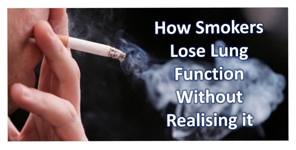
Damage from smoking can’t always be recognised by current lung function testing, this is because smoking causes 2 very distinct different conditions, Emphysema and/or Chronic Bronchitis.
Early stages of Emphysema are NOT detectible from normal lung function tests because it doesn’t affect your ability to breathe in, it only affects your ability to breathe out and it affects the air sacs at the end of the bronchial tubes.
Early stages of Bronchitis can be detected only because of the inflammation and mucus creation in the airways.
“Mucus dysfunction induced by cigarette smoke is complex and incompletely understood, but it involves adverse effects on the structure and function of cilia activation of ErbB receptors, decreased function of CFTR, and proinflammatory effects that increase mucin production while decreasing mucus hydration and clearance.”
The New England Journal of Medicine, 363(23), 2233-2247. 10. Fahy, J. V. & Dickey, B. F. (2010). Airway mucus function and dysfunction.
Damage to the lungs from smoking and pollution starts with one of the 2 following stages:
- Early Stage Emphysema
This is inflammation of the airway walls and the air sacs (aveoli), caused from breathing in the smoke and pollutants, this can lead to a condition called Air Trapping.
Air Trapping is when the airways leading to the air sacs become blocked up when you breath out, so the oxygen (O2) can go into the air sac, for gas exchange, but the carbon dioxide (CO2) can’t escape.
This Air Trapping (See Figure 1) can cause pressure build up in the air sacs similar to an expanding balloon which can damage the air sacs, leaving carbon particles (smoke) trapped in your lungs.
Or
- Early Stage Bronchitis and Emphysema
This is the inflammation of the airways and the air sacs which can lead to both Air Trapping and Collapsed Lung due to mucus build up and then mucus plugs in the airways leading to the air sacs.
Collapsed lungs are where the airways are fully closed due to the build-up of mucus in the airways, leading to a full plug where being stuck in the airways, stopping air from entering and exiting the air sac. Because air can’t escape from the air sac, the lungs absorb the CO2 from the air sac through the blood stream and move it to another air sac which can then release this gas. The air sac remains collapsed and is no longer used.
Medication is used to relieve inflammation, but it can’t release the trapped air or the mucus plug which causes the air sac to remain blocked.
If left untreated, this can lead to a decrease in lung volume of up to 33 millilitres every year. This is equivalent to more than 1 shot glass every year or over 4 shot glasses every 4 years.
So How Do You Remove the Mucus to help Improve Lung Capacity and Lung Function?
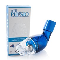
AirPhysio is an international award winning mechanical medical device which helps improve lung capacity and Lung function by doing the following:
- Mucus Removal and Clearance – The device shakes the airway walls (you can actually feel it working) and gets in behind the mucus and mucus plugs, helping to free it from the walls and start moving it up and out of the lungs to be swallowed or coughed out naturally.
- Lung Expansion – The device creates a positive pressure in the lungs and helps to open up closed and semi-closed airways, helping to restore lung capacity and maintain maximum lung capacity.
AirPhysio is an Australian Made and Owned Product, proudly partnered with Asthma Australia, is registered by the Therapeutic Goods Association (TGA) in Australia as a medical device and used in hospitals for patients with asthma to help asthmatics breathe easier, helping to improve effectiveness of medication when you need it most.
References
- World Health Organization (n.d.-a). Chronic obstructive pulmonary disease (COPD). Retrieved from http://www.who.int/respiratory/copd/en/
- World Health Organization (n.d.-b). Top 10 causes of death. Retrieved from http://www.who.int/mediacentre/factsheets/fs310/en/index2.html
- Rogers, D. F. (2007). Physiology of airway mucus secretion and pathophysiology of hypersecretion. Respiratory Care, 52(9) 1134-1149. Retrieved from http://rc.rcjournal.com/content/respcare/52/9/1134.full.pdf
- Rubin, B. K. (2014). Secretion properties, clearance, and therapy in airway disease. Published in Translational Respiratory Medicine, 109, 297-307. doi:10.1186/2213-0802-2-6
- Scheuch, G., Kohlhaufl, M., Moller, W., Brand, P., Meyer, T., Haussinger, K.,… Heyder, J. (2008). Particle clearance from the airways of subjects with bronchial hyperresponsiveness and with chronic obstructive pulmonary disease. Experimental Lung Research, 34, 531-549. doi:10.1080/01902140802341710
- Brown, J. S., Zeman, K. L. & Bennett, W. D. (2002). Ultrafine particle deposition and clearance in the healthy and obstructed lung. American Journal of Respiratory and Critical Care Medicine, 166, 1240–1247. doi:10.1164/rccm.200205-399OC
- Crotta, S., Ahlfors, H., Dingwell, K., & Smith, J. C. (2016). The aryl hydrocarbon receptor controls cyclin O to promote epithelial multiciliogenesis. Nature Communications, 1-11. doi:10.1038/ncomms12652
- Roth, M. (2015). Airway and lung remodelling in chronic pulmonary obstructive disease: A role for muscarinic receptor antagonists? Drugs, 75, 1-8. doi: 10.1007/s40265-014-0319-0
- Kulkarni, N., Pierse, N., Rushton, L., & Grigg, J. (2006). Carbon in airway macrophages and lung function in children. The New England Journal of Medicine, 355(1), 21-30
- Tashkin, D.P., Detels, R., Simmons, M., Liu, H., Coulson, A.H., Sayre, J., & Rokaw, S (1994). The UCLA population studies of chronic obstructive respiratory disease: XI. Impact of air pollution and smoking on annual change in forced expiratory volume in one second. American Journal Respiratory and Critical Care Medicine, 149(5), 1209-1217.
- Downs, S., Schindler, C., Liu, S., Keidel, D., Bayer-Oglesby, L., Brutsche, M.,… SAPALDIA Team (2007). Reduced exposure to PM10 and attenuated age-related decline in lung function. The New England Journal of Medicine, 357(23), 2338-2347. doi:10.1056/NEJMoa073625
- Ramos, F. L., Krahnke, J. S. & Kim, V. (2014). Clinical issues of mucus accumulation in COPD. International Journal of COPD, 9, 139-150. doi:10.2147/COPD.S38938
- Global Initiative for Chronic Obstructive Lung Disease, Inc. (2017). Pocket guide to COPD diagnosis, management, and prevention: A guide for health care professionals (2017 Ed.), 1-43. Retrieved from http://goldcopd.org/gold-reports/
- Medbø, A., & Melbye, H. (2008). What role may symptoms play in the diagnosis of airflow limitation?: A study in an elderly population. Scandinavian Journal of Primary Health Care, 26(2), 92–98. doi:10.1080/02813430802028938
- Vrijhoef, H.J., Diederiks, J.P., Wesseling, G.J., van Schayck, C.P., & Spreeuwenberg, C. (2003). Undiagnosed patients and patients at risk for COPD in primary health care: early detection with the support of non-physicians. Journal of Clinical Nursing, 12(3), 366-373. doi:10.1046/j.1365-2702.2003.00736.x
- Stratelis, G., Fransson, S., Schmekel, B., Jakobsson, P.,& Molstad, S. (2008). High prevalence of emphysema and its association with BMI: A study of smokers with normal spirometry. Scandinavian Journal of Primary Health Care, 26, 241-247. doi:10.1080/02813430802452732
- Stratelis, G., Jakobsson, P., Molstad, S., and Zetterstrom, O., (2004). Early detection of COPD in primary care: Screening by invitation of smokers aged 40 to 55 years. British Journal of General Practice, 54(500), 201-206.
- Giejer, R., Sachs, A., Verheij, T., Lammers, J., Salome, P., & Hoes, A. (2009). Are patient characteristics helpful in recognizing mild COPD (GOLD I) in daily practice? Scandinavian Journal of Primary Health Care, 24, 237-242. doi:10.1080/02813430601016894.
- Osadnik, C.R., McDonald, C.F., Jones, A.P. & Holland, A.E. (2012). Airway clearance techniques for chronic obstructive pulmonary disease (Review). Cochrane Database of Systematic Review, 3, 1-103. doi:10.1002/14651858.CD008328.pub2.
- Pryor, J. A. (1999). Physiotherapy for airway clearance in adults. European Respiratory Journal, 14, 1418-1424.
- Myers, T. (2007). Positive expiratory pressure and oscillatory positive expiratory pressure therapies. Respiratory Care, 52(10), 1308-1327.


