Contact dermatoscopy
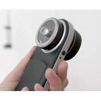
In essence, the dermatoscope consists of focusable magnification optics, an illumination source, and a transparent contact plate. Because most of the light that reaches the skin is reflected, a glass plate with a refractive index similar to that of the stratum corneum is used. The amount of light reflected is thereby considerably reduced. If additional fluid is applied to the skin lesion, the light can penetrate more deeply into the skin, and the epidermis essentially becomes transparent. Using the magnifying optics, the viewer can see details that are otherwise not visible to the naked eye.
Depending on the specification of the product, the contact fluid can either be disinfectant, immersion oil, or ultrasound gel. Disinfectants provide an additional microbicidal effect and are easy to handle. However, in order to prevent damage to the unit, the disinfectant should only be applied to the patient’s skin and not directly onto the contact plate. Because of its viscosity, immersion oil allows for extremely clear and distinct imaging. Ultrasound gel has advantages in the assessment of vascular structures because almost no pressure on the skin is required. The contact plate literally slides on the gel.
After the examination, the contact plate must be carefully wiped with a disinfected swab. Some dermatoscopes are also available with smaller contact plates. These have the advantage that structures such as the interdigital space, which are otherwise inaccessible, can be readily assessed.
Non-Contact dermatoscopy
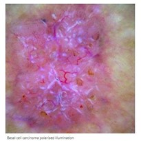
Dermatoscopes operating with polarised light instead of contact fluid have been available for several years. The polarised light reflected on the skin is suppressed by a polarisation filter. Using this technique, direct contact with the skin surface is not mandatory. The polarised dermatoscopes provide effects similar to those of contact dermatoscopy. Depending on the structures to be examined, there may be advantages or disadvantages; some structures are better identified with polarisation, while others are better identified without. For example, vascular patterns can be viewed more precisely with polarisation. However, the horn cysts of seborrheic keratosis are more prominent without polarisation.


